Breast cancers can be classified by different schemata. Every aspect influences treatment response and prognosis. Description of a breast cancer would optimally include multiple classification aspects, as well as other findings, such as signs found on physical exam. Classification aspects include stage (TNM), pathology, grade, receptor status, and the presence or absence of genes as determined by DNA testing:
Stage. The TNM classification for breast cancer is based on the size of the tumor (T), whether or not the tumor has spread to the lymph nodes (N) in the armpits, and whether the tumor has metastasized (M) (i.e. spread to a more distant part of the body). Larger size, nodal spread, and metastasis have a larger stage number and a worse prognosis.
The main stages are:
Stage 0 is a pre-cancerous or marker condition, either ductal carcinoma in situ (DCIS) or lobular carcinoma in situ (LCIS).
Stages 1–3 are defined as 'early' cancer with a good prognosis.
Stage 4 is defined as 'advanced' and/or 'metastatic' cancer with a poor prognosis.
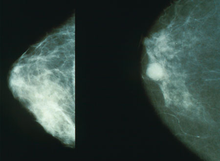
Histopathology. Breast cancer is usually classified primarily by its histological appearance. Most breast cancers are derived from the epithelium lining the ducts or lobules, and these cancers are classified as ductal or lobular carcinoma. Carcinoma in situ is growth of low grade cancerous or precancerous cells in particular tissue compartment such as the mammary duct without invasion of the surrounding tissue. In contrast, invasive carcinoma does not confine itself to the initial tissue compartment and invades the surrounding tissue.
Grade (Bloom-Richardson grade). When cells become differentiated, they take different shapes and forms to function as part of an organ. Cancerous cells lose that differentiation. In cancer grading, tumor cells are generally classified as well differentiated (low grade), moderately differentiated (intermediate grade), and poorly differentiated (high grade). Poorly differentiated cancers have a worse prognosis.
Receptor status. Cells have receptors on their surface and in their cytoplasm and nucleus. Chemical messengers such as hormones bind to these receptors, and this causes changes in the cell. Breast cancer cells may or may not have three important receptors: estrogen receptor (ER), progesterone receptor (PR), and HER2/neu.
ER+ cancer cells depend on estrogen for their growth, so they can be treated with drugs to block estrogen effects (e.g. tamoxifen), and generally have a better prognosis.
HER2+ breast cancer had a worse prognosis, but HER2+ cancer cells respond to drugs such as the monoclonal antibody, trastuzumab, (in combination with conventional chemotherapy) and this has improved the prognosis significantly. Cells with none of these receptors are called basal-like or triple negative.
DNA assays of various types including DNA microarrays have compared normal cells to breast cancer cells. The specific changes in a particular breast cancer can be used to classify the cancer in several ways, and may assist in choosing the most effective treatment for that DNA type.
Read More


